What is a CT Scan
Understanding the Technology, Benefits, and Risks of CT Scans for Medical Diagnosis
A CT scan, or computed tomography (CT) scan, is a type of imaging test that produces two-dimensional and three-dimensional pictures of the inside of an object. It uses X-rays in conjunction with computer technology to generate images. A doctor may order this procedure if they are looking for signs of tumors, infections or organ damage caused by diseases like cancer or heart failure.
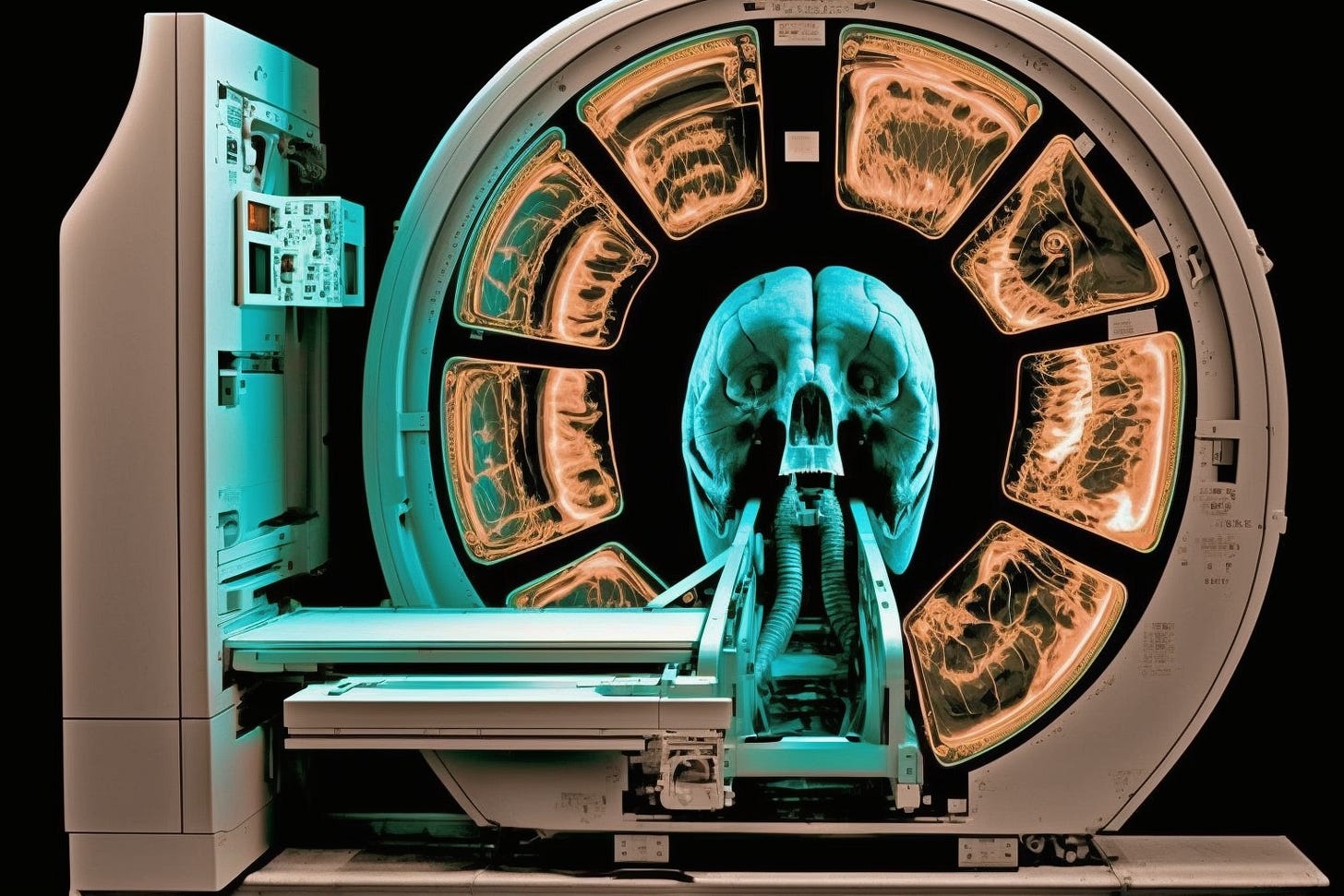
The actual process involves placing the patient on a table which moves through the center hole of a doughnut shaped scanner during testing time. Radiologists use powerful x-ray beams to create cross sectional scans throughout your body from all different angles and produce detailed 3D images that can be used for diagnosis purposes. The radiation exposure involved however, is minimal compared with other types of diagnostic tests because it focuses only on certain areas unlike full body radiographs.
Typically these exams take about 10 minutes but larger studies could range up to 1 hour depending upon complexity and size. Contrast agents such as iodine containing material might need to be injected intravenously prior to scanning so doctors have a clear view of highlighted structures. Thus it's important you notify personnel performing tests beforehand of any allergies or medications you are currently taking.
CT scans have become an invaluable tool not only in diagnosis but also when it comes to treatment planning as these images can help radiologists effectively pinpoint abnormalities within tissues before they start any form of surgical intervention. However, how does this particular technology compare with other scanning technologies?
The biggest advantage that CT Scanning has over other imaging techniques such as MRI (Magnetic Resonance Imaging), Ultrasound and X-Rays lies in speed – with some machines able to scan entire parts of the body within seconds compared to minutes for similar services on traditional devices. Additionally, unlike other methods which require careful positioning from experts; patients undergoing a CT scan just need to lie down and remain still while computer algorithms take care of constructing clear images from multiple angles rather than having expert hands guiding them into place every time. This means that clearer visuals are provided without compromising patient safety or comfort during examinations. Although radiation exposure levels remain low, so far there hasn't been any major long lasting side effects linked directly to related usage, however further studies may be needed eventually should new advancements see higher doses being emitted by modern scanners on occasion.
Another benefit offered by using CT scans instead of alternatives include greater detail resolution, allowing smaller structures deep inside the subject's bodies to be made visible even if they're made out of bone marrow etc, something conventional ultrasonic based instruments tend to struggle to pick up accurately depending on the environment where the test was conducted. Lastly, cost factors must be taken into account here too considering the level of sophistication involved in the construction process, setup, and maintenance – both more expensive, yet potential benefits outweigh drawbacks overall proving a great addition to an existing suite of diagnostic procedures already available today.
All things considered then CT scans stand head and shoulders above the rest in terms of what they offer. They can provide a versatile range of capabilities that are useful in a variety of applications ranging from simple illnesses to complex situations.
A CT scanner consists of several key components that all contribute towards producing high quality diagnostic images. The main parts of a typical system include:
X-ray tube: This is responsible for generating x-rays that pass through the patient’s body being scanned as well as collimating them into fan beams directed at detectors located behind the table on which they lie down during their examination.
Gantry: This is where all other major elements such as detector arrays, electronics cabinets etc., are mounted; it rotates around its vertical axis while performing image acquisition over 360 degrees within only few seconds.
Data Acquisition System (DAS): It collects raw data produced by electronic circuits connected between each array element via cables running from gantry assembly to consoles outside the room itself.
Reconstruction Computer Software Package: Using information gathered from DAS along with algorithms, specific reconstructions take place before final results become available electronically/digitally.
Apart from above-mentioned hardware devices, many software programs help create 3D models out of 2D slices taken using different projections allowing physicians to obtain a more comprehensive view about inner structures and organs than ever before. The latest developments focus on improving speed and accuracy of analyses made in the process.


